February 27, 2026
From letter to structured care data
Stijn Bruggeman
The problem
A general practitioner receives on average 10 to 30 letters per day: discharge summaries, specialist reports, imaging results. These documents contain crucial information about diagnoses, medication changes and treatment plans.
Yet they end up in the EHR as unstructured attachments: files with no structure whatsoever. The information is buried in running text. To update the patient record, the GP must read each letter in full, interpret the relevant data and enter it manually.
The result: information gets lost, records are incomplete, and follow-up care is compromised. Not because the information isn’t there, but because it is not accessible.
What if every new diagnosis, medication change and treatment plan would become visible automatically? Let’s walk through it step by step, using a real discharge letter as an example.
The letter
A 74-year-old patient was admitted for an RSV infection. Below is an example of a typical discharge letter: several paragraphs of text containing diagnoses, examinations, treatments and medication.
Dear colleague,
Verlinden, Jan Pieter - born 17-04-1948, Molenstraat 23/0101, 2170 Antwerp - was admitted from 06-01-2023 to 09-01-2023 at the Department of Pulmonology in ZNA Stuivenberg for RSV infection.
Reason for admission / Referral
Pneumonia
History
Several days of malaise, anorexia, fatigue, muscle pain, last two days fever and increasing dyspnea and cough.
Visited the GP, yesterday COVID swab negative.
GP started inhaler, no improvement after which referred to the emergency department.
Substance use
The patient has never smoked.
The patient does not drink alcohol.
The patient does not use drugs.
Allergies
None.
Social history
Lives at home with his wife. They still manage all household tasks independently.
Active isolation indication
08/01/2023 Respiratory syncytial virus (RSV), isolation type: Contact - droplet isolation
Physical examination
General: still vital man for his age, T 37.1°C
Heart: normal heart sounds
Lungs: bilateral vesicular breath sounds, bibasal fine crackles L>R, no wheezing
Skin: no infection
Neuro: no abnormalities
Additional investigations
ABG 06/01/23: pH 7.47, pCO2 34.0, pO2 56.3, Na 133
Lab 06/01/23: Hb 10.3, WBC normal, left shift, CK 219, troponins 74.1 (non-evolving), CRP 134.5
PCR influenza/COVID19: Negative
Respiratory panel 06/01/23: RSV positive
Blood cultures 07/01/23: negative
Chest X-ray 06/01/23:
Post-sternotomy status.
Heart and mediastinal width within normal limits. Aortic knob atheromatosis. Satisfactory transparency of the lung fields. Discrete increased markings in the infrahilar regions and retrocardiac suggestive of chronic inflammation/bronchitis. The costophrenic angles appear clear. No perihilar congestion signs. Increased kyphosis of the thoracic spine.
CT pulmonary embolism 06/01/23:
Conclusion:
No evidence of pulmonary embolism. Bronchiectatic airways in the right middle lobe and both lower lobes. Peribronchovascular ground-glass opacity in both lower lobes and dorsobasal in the left lower lobe suggestive of inflammatory pathology. Also patchy opacities peribronchovascular in the right upper lobe also suggestive of underlying inflammation.
Conclusion
Your 74-year-old patient was admitted to the pulmonology department from 06/01/23 to 09/01/2023. We retain:
1. RSV infection with type 1 respiratory insufficiency
Presentation at the emergency department with several days of general malaise with fever and cough with progressive dyspnea. Bibasal fine crackles on lung auscultation. Biochemically we retain an inflammatory blood panel with elevated CRP. Respiratory panel positive for RSV. Blood cultures negative. On chest X-ray we see signs of chronic inflammation/bronchitis with on CT thorax bronchiectasis and patchy opacities peribronchovascular in the right upper lobe. At admission empirical Augmentin was started, this is continued after discharge so that the total treatment duration is 7 days. During admission oxygen dependency for which 3L O2/min was started, this could be tapered guided by saturation and clinical status. There is no oxygen dependency at discharge.
2. Diabetes type 2
For which treatment with Komboglyze 2.5/850mg twice daily. Since Komboglyze has been withdrawn from the market, this was changed to Sitagliptin/Metformin 2.5/850mg twice daily.
3. Type 2 ischemia
Post-CABG in 2018. Recent (in 03/2022) admission to cardiology for NSTEMI. Coronary angiography showed new severe stenosis at the mid-LAD proximal to the LIMA anastomosis. Conservative management with Brilique for 1 year.
Troponins at admission elevated to 75 ng/L without complaint of retrosternal pain. TTE during admission shows no new contractility abnormalities. Thus suspected type 2 ischemia secondary to item 1.
4. Arterial Hypertension
During admission consistently high blood pressures around 160/90mmHg. For this, Amlor 5 mg once daily was started. Please follow up.
Current medication: home medication
Saxagliptin 2.5 mg + metformin hydrochloride 850 mg tablet (oral); oral; twice daily 1 tablet with meals
Amlodipine 5 mg tablet (oral); oral; once daily 1 tablet
Ticagrelor 90 mg tablet (oral); oral; twice daily 1 tablet
Pantoprazole 20 mg gastro-resistant tablet (oral); oral; once daily 1 tablet on empty stomach
Ezetimibe 10 mg + rosuvastatin 40 mg tablet (oral); oral; once daily 1 tablet
Acetylsalicylic acid 80 mg gastro-resistant tablet (oral) (cardiovasc.); oral; once daily 1 tablet
With collegial regards,
dr. Pieter Peeters
On behalf of
dr. Katrien De Smet
This letter has been electronically validated and is therefore not signed.
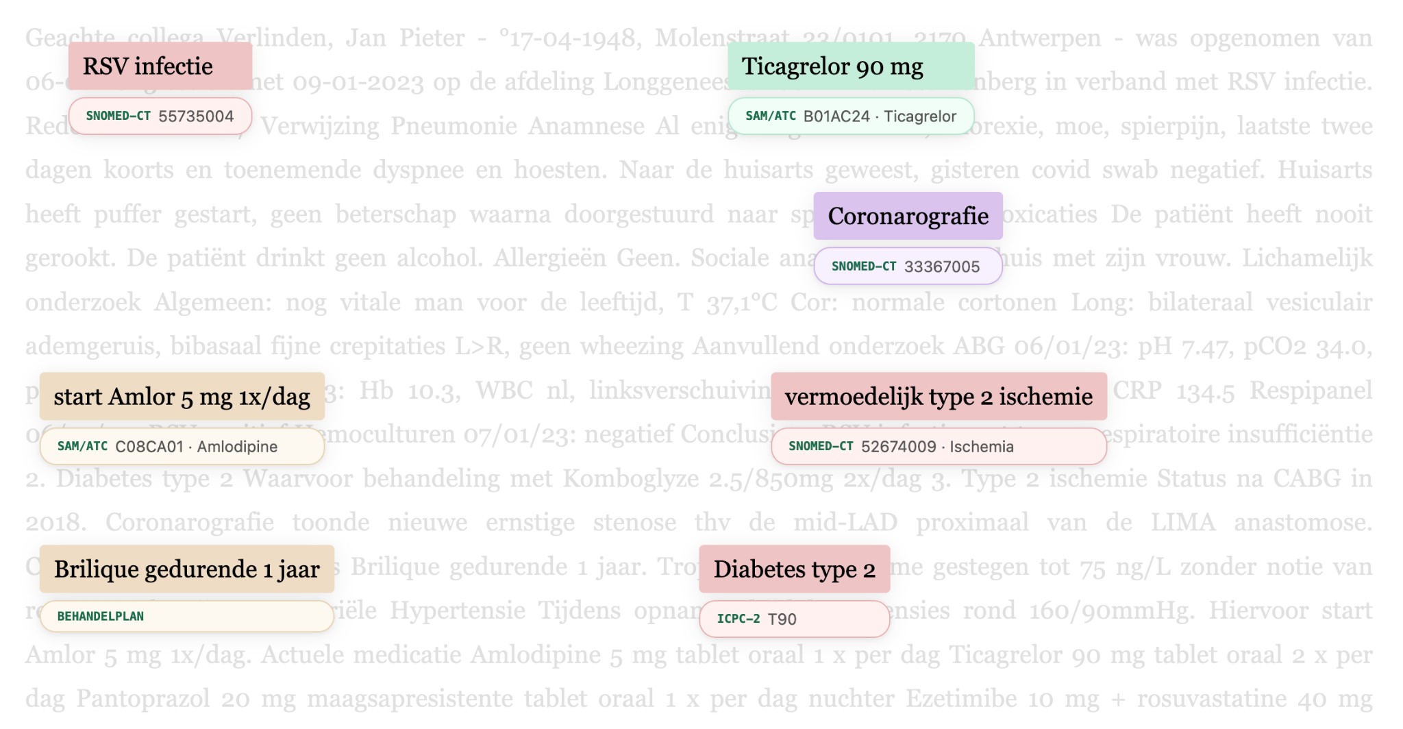
Step 1: Recognition
The system reads the entire letter and marks every clinically relevant fragment. Each fragment is assigned a type: diagnosis, procedure, care plan, medication, lab result, and more. Because these categories align with the FHIR standard, the data is universally interoperable with other healthcare systems.
In this letter, more than 100 fragments were automatically recognized, from the primary diagnosis “RSV infection” to individual lab values such as “CRP 134.5”.
Dear colleague,
Verlinden, Jan Pieter - born 17-04-1948, Molenstraat 23/0101, 2170 Antwerp - was admitted from 06-01-2023 to 09-01-2023 at the Department of Pulmonology in ZNA Stuivenberg for RSV infection.
Reason for admission / Referral
Pneumonia
History
Several days of malaise, anorexia, fatigue, muscle pain, last two days fever and increasing dyspnea and cough.
Visited the GP, yesterday COVID swab negative.
GP started inhaler, no improvement after which referred to the emergency department.
Substance use
The patient has never smoked.
The patient does not drink alcohol.
The patient does not use drugs.
Allergies
None.
Social history
Lives at home with his wife. They still manage all household tasks independently.
Active isolation indication
08/01/2023 Respiratory syncytial virus (RSV), isolation type: Contact - droplet isolation
Physical examination
General: still vital man for his age, T 37.1°C
Heart: normal heart sounds
Lungs: bilateral vesicular breath sounds, bibasal fine crackles L>R, no wheezing
Skin: no infection
Neuro: no abnormalities
Additional investigations
ABG 06/01/23: pH 7.47, pCO2 34.0, pO2 56.3, Na 133
Lab 06/01/23: Hb 10.3, WBC normal, left shift, CK 219, troponins 74.1 (non-evolving), CRP 134.5
PCR influenza/COVID19: Negative
Respiratory panel 06/01/23: RSV positive
Blood cultures 07/01/23: negative
Chest X-ray 06/01/23:
Post-sternotomy status.
Heart and mediastinal width within normal limits. Aortic knob atheromatosis. Satisfactory transparency of the lung fields. Discrete increased markings in the infrahilar regions and retrocardiac suggestive of chronic inflammation/bronchitis. The costophrenic angles appear clear. No perihilar congestion signs. Increased kyphosis of the thoracic spine.
CT pulmonary embolism 06/01/23:
Conclusion:
No evidence of pulmonary embolism. Bronchiectatic airways in the right middle lobe and both lower lobes. Peribronchovascular ground-glass opacity in both lower lobes and dorsobasal in the left lower lobe suggestive of inflammatory pathology. Also patchy opacities peribronchovascular in the right upper lobe also suggestive of underlying inflammation.
Conclusion
Your 74-year-old patient was admitted to the pulmonology department from 06/01/23 to 09/01/2023. We retain:
1. RSV infection with type 1 respiratory insufficiency
Presentation at the emergency department with several days of general malaise with fever and cough with progressive dyspnea. Bibasal fine crackles on lung auscultation. Biochemically we retain an inflammatory blood panel with elevated CRP. Respiratory panel positive for RSV. Blood cultures negative. On chest X-ray we see signs of chronic inflammation/bronchitis with on CT thorax bronchiectasis and patchy opacities peribronchovascular in the right upper lobe. At admission empirical Augmentin was started, this is continued after discharge so that the total treatment duration is 7 days. During admission oxygen dependency for which 3L O2/min was started, this could be tapered guided by saturation and clinical status. There is no oxygen dependency at discharge.
2. Diabetes type 2
For which treatment with Komboglyze 2.5/850mg twice daily. Since Komboglyze has been withdrawn from the market, this was changed to Sitagliptin/Metformin 2.5/850mg twice daily.
3. Type 2 ischemia
Post-CABG in 2018. Recent (in 03/2022) admission to cardiology for NSTEMI. Coronary angiography showed new severe stenosis at the mid-LAD proximal to the LIMA anastomosis. Conservative management with Brilique for 1 year.
Troponins at admission elevated to 75 ng/L without complaint of retrosternal pain. TTE during admission shows no new contractility abnormalities. Thus suspected type 2 ischemia secondary to item 1.
4. Arterial Hypertension
During admission consistently high blood pressures around 160/90mmHg. For this, Amlor 5 mg once daily was started. Please follow up.
Current medication: home medication
Saxagliptin 2.5 mg + metformin hydrochloride 850 mg tablet (oral); oral; twice daily 1 tablet with meals
Amlodipine 5 mg tablet (oral); oral; once daily 1 tablet
Ticagrelor 90 mg tablet (oral); oral; twice daily 1 tablet
Pantoprazole 20 mg gastro-resistant tablet (oral); oral; once daily 1 tablet on empty stomach
Ezetimibe 10 mg + rosuvastatin 40 mg tablet (oral); oral; once daily 1 tablet
Acetylsalicylic acid 80 mg gastro-resistant tablet (oral) (cardiovasc.); oral; once daily 1 tablet
With collegial regards,
dr. Pieter Peeters
On behalf of
dr. Katrien De Smet
This letter has been electronically validated and is therefore not signed.
Step 2: Normalization
Recognition alone is not enough. Free text is difficult to search and hard to compare across systems. That is why each recognized fragment is linked to standardized medical codes, such as SNOMED-CT for clinical concepts and ICPC-2 for primary care, as well as LOINC and other international terminologies.
This way, “RSV infection” is no longer just a highlighted piece of text, but a coded concept that any healthcare system can understand, regardless of how the specialist originally phrased it.
Newly initiated medication. The GP needs to know this for follow-up.
Ongoing treatment plan with a specific duration. Essential for medication follow-up.
Even uncertain findings are recognized and correctly classified.
This makes the information searchable, comparable and reusable. Not only within the practice’s own EHR, but also for data exchange with other healthcare systems through standards like FHIR.
Step 3: Comparison with the patient record
The structured data from the letter is automatically compared with the existing patient record, which is structured in the same way. What is already known? What is new?
The GP no longer needs to piece it together: the system directly shows which information requires attention.
New for this patient
- RSV infectionnew diagnosis
- Amlor 5 mg once dailynew medication
- Suspected type 2 ischemianew finding
Already known in the record
- Diabetes type 2existing diagnosis
- Post-CABG (2018)known history
- Saxagliptinongoing medication
The result
Instead of a lengthy attachment, the GP immediately sees what is relevant and new: which diagnoses, which medication, which treatment plan. Without having to read the entire letter.
Reading through letters, identifying new data that is not yet in the record and manually updating it: this is an enormous administrative burden that recurs daily. By automating this processing, the physician not only saves time, but can focus on what truly matters: staying informed about the important updates for their patients.
Looking ahead
When all incoming information is captured in a structured and coded way, it opens the door to population-level analytics.
How many patients in the practice have diabetes and qualify for a care pathway? Which patients with chronic kidney disease need follow-up?
Quality indicators and population management become possible, without any additional registration burden for the GP. The data is already there. It just needs to be unlocked.
Want to explore Co-Medic further?
Connect with our team or keep reading more insights from our blog archive.
Available in:
Related articles
Mar 25, 2026
Introducing Co-Medic Research: Identify patient populations in primary care
A new tool for identifying patient populations in primary care using structured clinical data, now in private beta with select partners.
Read more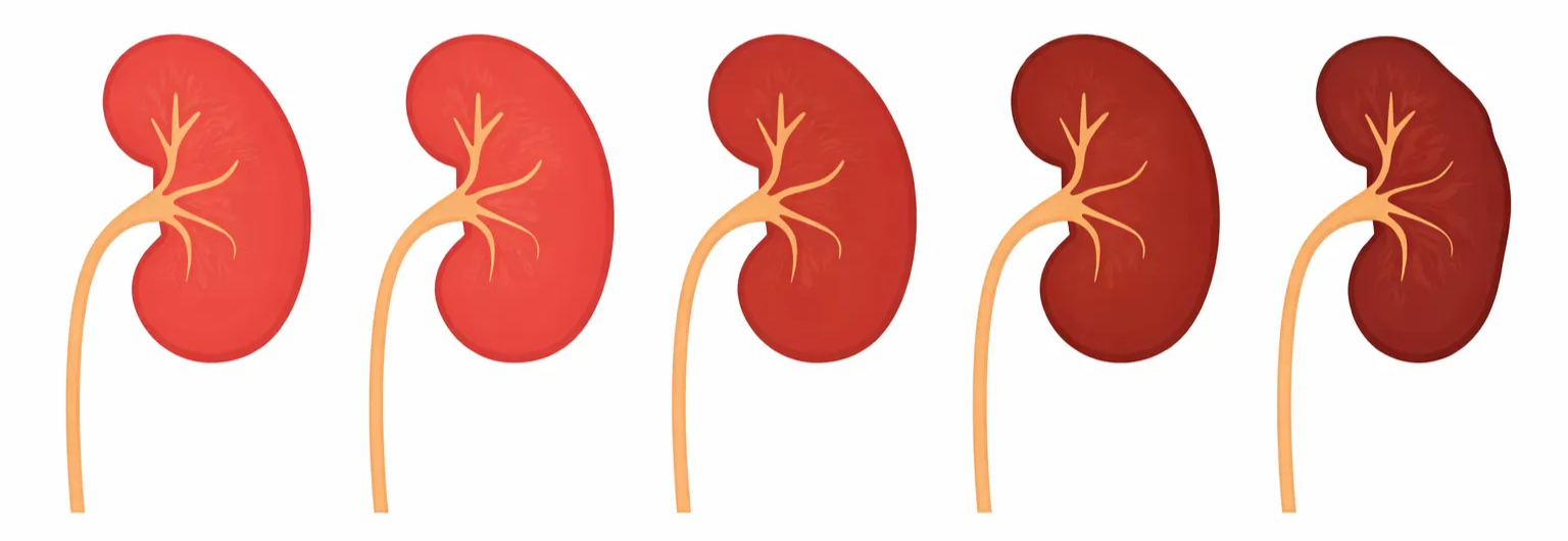
Feb 5, 2026
Chronic kidney disease: from underdiagnosis to overview with population management
CKD is often insufficiently followed up in primary care. Population management helps identify and follow patients according to guidelines.
Read more
Jan 27, 2026
How Co-Chat relieves daily administrative tasks
Dr. Bram Spinnewijn shows how he writes referral letters during consultations with Co-Chat, without GDPR concerns.
Read more